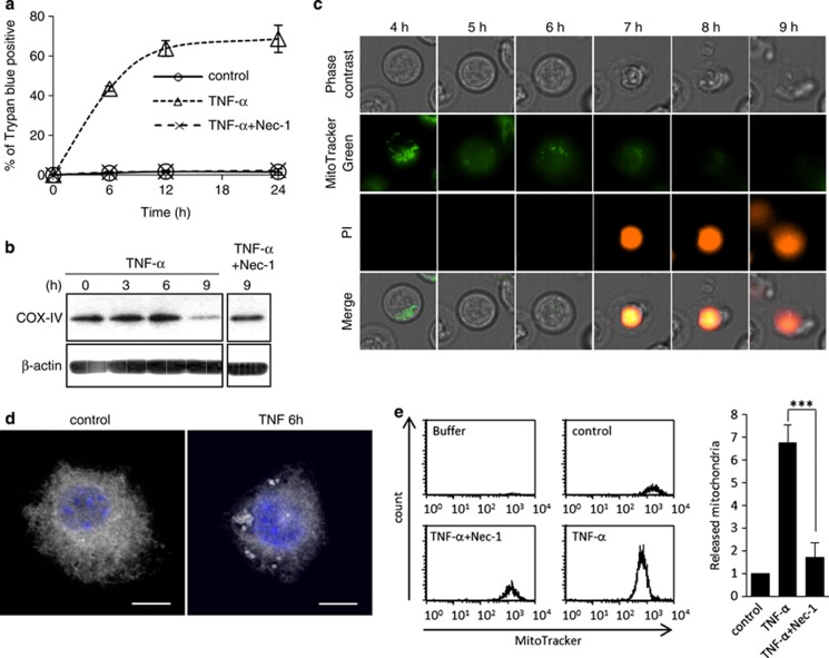
Figure 3.
TNF-α induces early release of mitochondria during necroptosis. (a) FADD-deficient Jurkat cells were treated with 10 ng/ml of TNF-α with/without 40 μM Nec-1, and stained with trypan blue. Data shown are from three independent experiments. (b) FADD-deficient Jurkat cells were treated with 10 ng/ml of TNF-α with/without 40 μM Nec-1 for the indicated time and cell lysates were blotted with anti-COX-IV antibody. β-actin antibody is used as loading control. (c) Time-lapse imaging of TNF-α-treated FADD-deficient Jurkat cells. Cells were treated with 10 ng/ml TNF-α in the presence of MitoTracker Green and propidium iodide (red). (d) Fluorescence microscopic images of L929 cells treated or not with 5 ng/ml of TNF-α for 6 h. Cells were stained with MitoTracker Deep Red and DAPI to visualize the cell nucleus. Scale bars: 10 μm. (e) The pellets were collected from the supernatant of the FADD-deficient Jurkat cells cultured medium after 24 h incubation with 10 ng/ml of TNF-α with/without 40 μM Nec-1, and the crude pellet was stained with MitoTracker Green. The fluorescence-labeled pellets were analyzed by flow cytometry. The graph shows the fold increase of MitoTracker-positive events when compared with the control. Data shown are mean values±S.E.M. of three independent experiments. ***P<0.001 by the Student's t-test

