Figure 5.
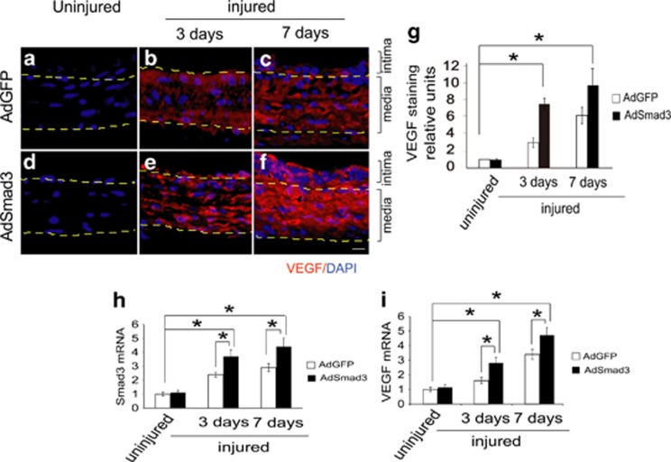
Smad3 stimulates VEGF expression in balloon-injured rat carotid arteries. (a–f) Effect of Smad3 up-regulation on VEGF expression in balloon-injured rat carotid arteries. Sham surgery (a and d) or balloon angioplasty (b, c, e and f) was performed in rat common carotid arteries followed by infusion (200 μl virus, 2.5 × 10⁹ PFUs/ml) of AdGFP or AdSmad3 for 20 min. Arteries were retrieved 3 days (b and e) or 7 days (c and f) after injury and immunostaining of VEGF-164 (red) was performed on carotid sections as described in the Materials and Methods. Shown are representative images from a total of four animals. Dashed lines define the media layer. (g) Quantification of protein expression levels of VEGF in experiments (a–f) based on relative fluorescence intensity (n=4; *P<0.05 compared between injured and uninjured arteries). (h and i) Quantification of mRNA levels of Smad3 (h) and VEGF (i) in rat carotid arteries. In parallel to experiments (a–f), rat carotid arteries at 3 and 7 days were homogenized for mRNA extraction and RT-PCR was used to quantify levels of Smad3 and VEGF-164 (shown in h and i, respectively). Each bar represents mean±S.E.M. of four animals (*P<0.05 compared between injured and uninjured arteries, or without and with Smad3 overexpression)

