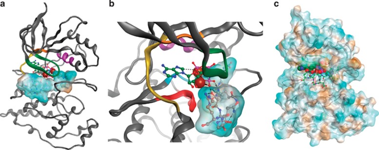
Figure 6.
Theoretical molecular model of JNK2 substrate recognition of the Bid peptide. (a) Ribbon diagram of JNK2-ANP homology model with docked Bid peptide. The Bid peptide is rendered as a hydrophobic surface with brown being the most hydrophobic and blue being hydrophilic. Ribbon is colored according to kinase domains with green as the G-loop, HRD in red, hinge in yellow and alphaC in purple. (b) Close-up view of (a) highlighting the bonding of ANP with the Bid peptide. (c) JNK2 surface rendering colored according to hydrophobicity (the Bid peptide, sticks; ANP space fill mode)

