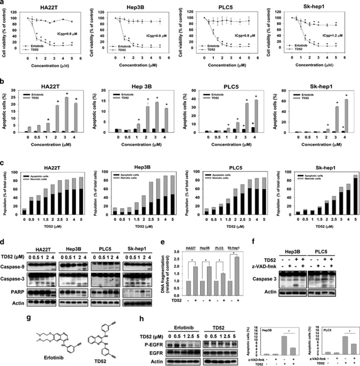
Figure 1.
TD52, a derivative of erlotinib, showed a better pro-apoptotic effect of HCC cells than erlotinib. (a) Pro-apoptotic effects of erlotinib and TD52 in HCC cells. HCC cells were exposed to erlotinib and TD52 at the indicated concentrations for 48 h, and then measured by MTT assay. IC50 of TD52 in different cells were illustrated. Points, mean; bars, S.D. (n=3). (b) Dose-dependent effect of TD52 and erlotinib on cell apoptosis in HCC cells. Percentage of apoptotic cells of HCC cells were measured by flow cytometry (sub-G1) after exposure to erlotinib and TD52 at indicated concentrations for 24 h. Bar, mean; error bars, S.D. (n=3). (c–e) Dose-escalation effect of TD52 in four different HCC cell lines. After incubation with TD52 at the indicated concentration in DMEM with 5% FBS for 24 h (for western blot and DNA fragmentation test) and 48 h (for annexin-V/PI double-staining assay), the HCC cells were harvested and analyzed by annexin-V/PI double-staining assay (c), western blot for the expression of caspases-3, caspases-9 and PARP (d), and DNA fragmentation test (e, n=6). Data of western blotting are representative of three independent experiments. Analysis of HCC cell condition in the annexin-V/PI double-staining assay was performed by flow cytometry and the proportion of apoptotic cells was determined as the sum of pro-apoptotic cell plus late apoptotic cell (annexin-V-FITC-positive cells). Result of flow cytometry was detailed in Supplementary Figure 1. (n=3). (f) The pro-apoptotic effects induced by TD52 was diminished by co-treatment with z-VAD-fmk. HCC cells were treated with TD52 (2 μM) with or without the pan-caspase inhibitor, z-VAD-fmk (100μM), for 48 h, and then cells were analyzed by western blot (upper panel) and flow cytometry (lower panel). (g) The structure of erlotinib (upper) and TD52 (lower). (h) TD52 exhibit little EGFR phosphorylation activity. HCC cells treated with erlotinib or TD52 for 24 h and EGFR phosphorylation activity was determined by western blotting

