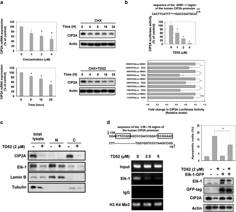
Figure 3.
TD52 downregulated transcription of CIP2A via interfering Elk-1 function. (a) TD52 affected transcription of CIP2A in a dose- and time-dependent manner. Left panel, PLC5 cells were treated with D52 at indicated doses (upper panel) and time (lower panel); CIP2A mRNA was quantified by reverse transcription-PCR. Bar, mean; error bars, S.D. (n=3) Right panel, after treating cells with 100 mg/ml CHX in the presence (lower) or absence (upper) of TD52 for the indicated length of time, the expression of CIP2A protein in whole-cell lysates was assessed by western blot. (b) Identification of the CIP2A proximal promoter region that was affected by TD52. CIP2A promoter activities at different doses of TD52 were first examined (upper panel). Promoters with five different length of deletion were constructed as detailed in the ‘Materials and methods' section. Followed by transfecting with the mutant clone or the wide-type promoter for 24 h, PLC5 cells were subsequently exposed to TD52 at 2μM and assayed for luciferase activity after another 24 h. (n=3, *P<0.05, Bar, mean; error bars, S.D. NS, nonsignificant). (c) TD52 affected the protein expression of Elk-1 and CIP2A in PLC5 cells. After exposure with TD52 at 2 μM for 24 h, PLC5 cells were lysed and whole-cell extract, cytoplasmic and nuclear fractions were prepared. The expression of CIP2A and Elk-1 were analyzed by western blot. Tubulin and lamin B were used as loading control. (d) Inhibition of the binding of Elk-1 to the CIP2A promoter determined the effect of TD52 in PLC5 cells. Left panel, a ChIP assay was performed on TD52- or mock-treated PLC5 cells. The representative sequence of the CIP2A promoter (upper), □ indicated the putative binding sites of Elk-1. ChIP assay was conducted to evaluate the DNA-binding ability of Elk-1 on the putative binding sites of CIP2A promoter region (lower). PLC5 cells were treated with TD52 at the indicated concentrations for 16 h and fixed for ChIP assay. The products were amplified by PCR and the result was presented by gel-electrophoresis. Right panel, ectopic expression of Elk-1 reduced the pro-apoptotic effects of TD52 in PLC5 cells. Cells were transfected with Elk-1-GFP for 48 h and exposed to TD52 (2 μM) for 24 h. Bar, mean; error bars, S.D. (n=3)

