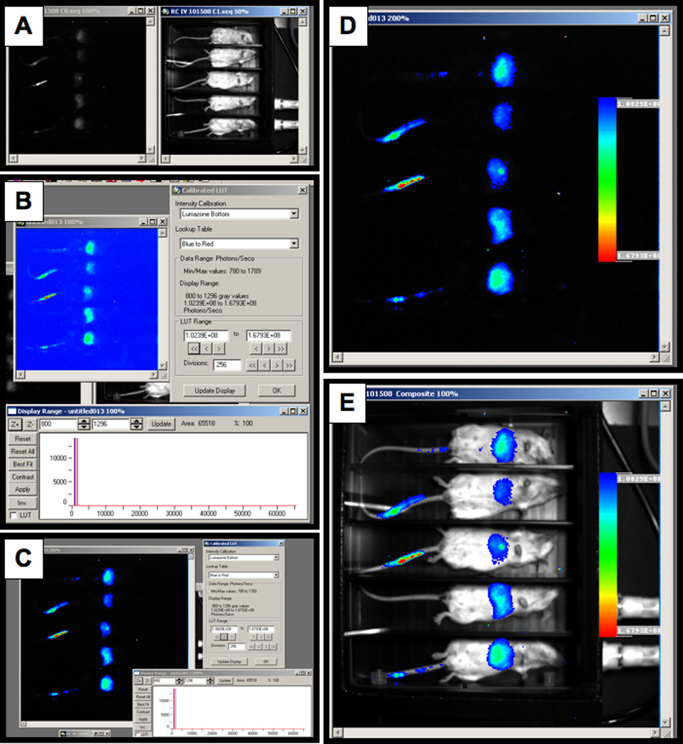
Figure 1. Bright light and chemiluminescent images.

A) Image of the light emitted in vivo and a white light image of the animals. B) Calibrated LUT function. C) Best Fit in the display range window to remove low level background signal. D) Background area selection. E) Image overlay selection.
