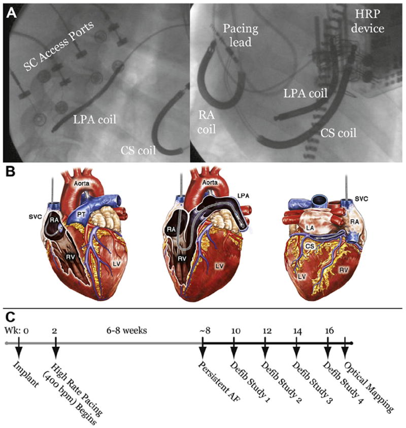
Figure 1. Chronically Implanted Lead Positions and Experimental Timeline.

(A) Fluoroscopic images of the anatomic positions of chronically-implanted transvenous leads and subcutaneous (SC) access ports from left lateral (left panel) and left anterior oblique (right panel) views. (B) Schematic depiction of lead positions. Shocks were delivered from the right atrium (RA) coil to the left pulmonary artery (LPA) coil or the RA coil to the coronary sinus (CS) coil. (C) Experimental timeline showing model development and approximate times of defibrillation (defib) studies. AF = atrial fibrillation; HRP = high rate pacing; LA = left atrium; LV = left ventricle; PT = pulmonary trunk; RV = right ventricle; SVC = superior vena cava; Wk = week.
