
Figure 1.
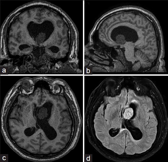
(a) T1-weighted coronal view. Hypointense nodular image in the floor of the left lateral ventricle, producing mild-moderate, noncommunicating hydrocephalus. (b) T1-weighted sagittal view. Note the involvement of the brainstem, which explains the symptoms of the patient. (c) T1-weighted axial view showing a nodular, hypointense lesion with small cystic areas in its interior. (d) Axial view, FLAIR. Note the absence of perilesional edema, and the lack of contrast enhancement