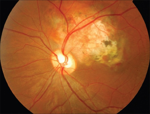
Figure 4.

Left peripapillary extrafoveal osteoma of the choroid measures 3 by 2 disc diameters and is associated with subfoveal CNV that responded to 2 consecutive intravitreal bevacizumab injections with visual improvement over one year of follow-up. B-scan, OCT and fluorescein angiography transits are shown in Figures 7-9 (Courtesy of Dr Eman Al Kahtani, King Khaled Eye Specialist Hospital)
