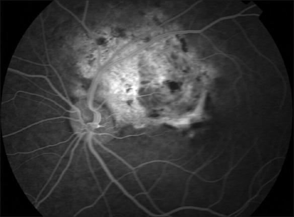
Figure 6.

Fluorescein angiography 35-second transit reveals dye leakage in the foveal region (CNV) and dye staining in the area of chronic RPE decompensation overlying the choroidal osteoma at 1 o'clock near the peripapillary area (Courtesy of Dr Eman Al Kahtani, King Khaled Eye Specialist Hospital)
