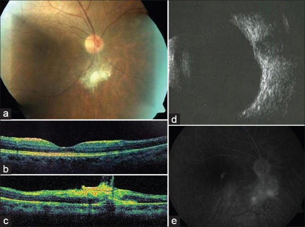
Figure 3.

2 years after photodynamic therapy: (a) Marked regression of the tumor. (b) Optical coherence tomography (OCT)(fovea) shows disappearance of the serous retinal detachment. (c) OCT (lesion) precisely demonstrates the intraretinal location of the tumor with overlying epiretinal membrane. (d) Ultrasonography discloses minimal elevation due to residual scar. (e) Late phase angiography showing hyperfluorescence with staining of the residual, intraretinal fibrotic tissue
