Figure 1.
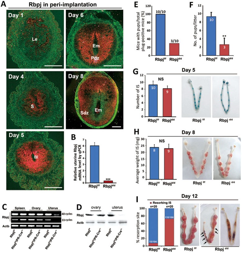
Rbpj is expressed in a spatiotemporal manner in the uterus and is critical for normal pregnancy. (A) In situ hybridization showing the spatiotemporal expression of Rbpj in WT uteri on days 1, 4, 5, 6 and 8 of pregnancy. Bl, blastocyst; Le, luminal epithelium; Pdz, primary decidual zone; Sdz, secondary decidual zone; S, stroma. (B) Real-time quantitative PCR analysis of uterine Rbpj mRNA in Rbpjf/f and Rbpjd/d mice. Data shown represent the mean ± SEM. ***P < 0.01. (C) RT-PCR analysis of Rbpj expression in the spleen, ovary and uterine stromal cells of Rbpjf/f and Rbpjd/d mice. Rbpj mRNA expression was efficiently deleted in the uteri of the Rbpjd/d mice but still abundant in the spleen and ovary. (D) Immunoblotting analysis of Rbpj protein in the ovaries and uteri on day 4 of pregnancy dissected from Rbpjf/f and Rbpjd/d mice. (E) Pregnancy outcomes in Rbpjf/f and Rbpjd/d mice. (F) Average litter sizes in Rbpjf/f and Rbpjd/d mice. **P < 0.01. (G) Morphologically normal implantation in Rbpjd/d mice compared with Rbpjf/f mice as determined by blue dye injection on day 5. The average number of implantation sites is comparable between the Rbpjf/f and Rbpjd/d mice. IS, implantation site; NS, not significant. (H) The weight of the implantation sites and representative uteri from Rbpjf/f and Rbpjd/d females on day 8 of implantation. IS, implantation site; NS, not significant. (I) Resorption rate and representative uteri from Rbpjf/f and Rbpjd/d females on day 12. The black arrowheads denote the resorption sites. In I, the numbers within bars indicate the number of resorption events divided by the total number of implantation sites. In F-H, the numbers within bars indicate number of females examined for each group.

