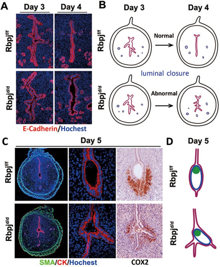
Figure 3.
Rbpjd/d females show defective luminal closure and deflected embryo orientation at the time of initial implantation. (A) Abnormal luminal closure and increased epithelial branching in Rbpjd/d females evidenced by the immunofluorescence of E-cadherin in day 3 and day 4 uteri. Cy3-labeled E-cadherin is shown in red; Hoechst 33342-labeled nuclei are shown in blue. (B) Diagram illustrating the process of luminal closure from day 3 to day 4 in Rbpjf/f and Rbpjd/d uteri. (C) Co-immunofluorescence of CK and SMA and the immunohistochemistry of COX2 in Rbpjf/f and Rbpjd/d implantation sites on day 5. SMA, smooth muscle actin; CK, cytokeratin. (D) Diagram of embryo implantation in day 5 implantation sites in Rbpjf/f and Rbpjd/d mice.

