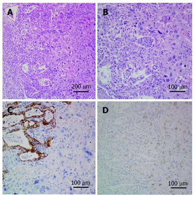
Figure 6.

Microscopic specimen. A and B: Both adenocarcinoma and squamous cell carcinoma components. Adenocarcinoma component shows gland formation and squamous carcinoma component shows keratinization (hematoxylin and eosin staining, A: × 10 magnification; B: × 20 magnification); C: Immunohistochemical (IHC) staining for cytokeratin (low), on the left side adenocarcinoma is strongly positive and on the right side squamous cell carcinoma is negative (IHC × 20 magnification); D: IHC staining for P63, on the left side adenocarcinoma is negative and on the right side squamous cell carcinoma is nuclear positive (IHC × 20 magnification).
