Figure 3.
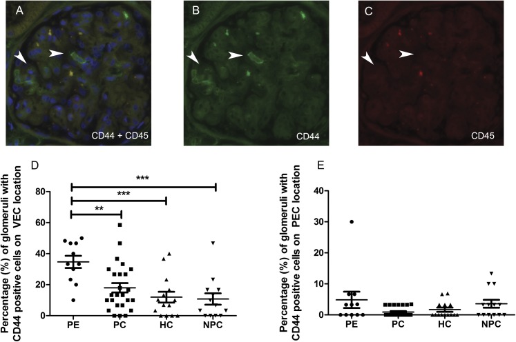
Double staining of CD44 and CD 45. Sections were costained for CD44 (green) and CD45 (red), and the number of CD44-positive/CD45-negative cells was scored within the glomeruli. (A) Double staining of CD44-positive/CD45-negative cells on a podocyte location (arrowheads). The nuclei were counterstained with 4′,6-diamidino-2-phenylindole (blue). (B and C) Note that the CD44-positive cells (B) are CD45-negative (C). (D) The number of CD44-positive cells on a podocyte (visceral epithelial cell) location (**P=0.01; ***P=0.001). (E) The number of CD44-positive cells on a parietal epithelial cell location (P=0.07). In D and E, each symbol represents an individual patient or control. The lines represent the mean value, and the error bars represent the SD. PEC, parietal epithelial cell; VEC, visceral epithelial cell.

