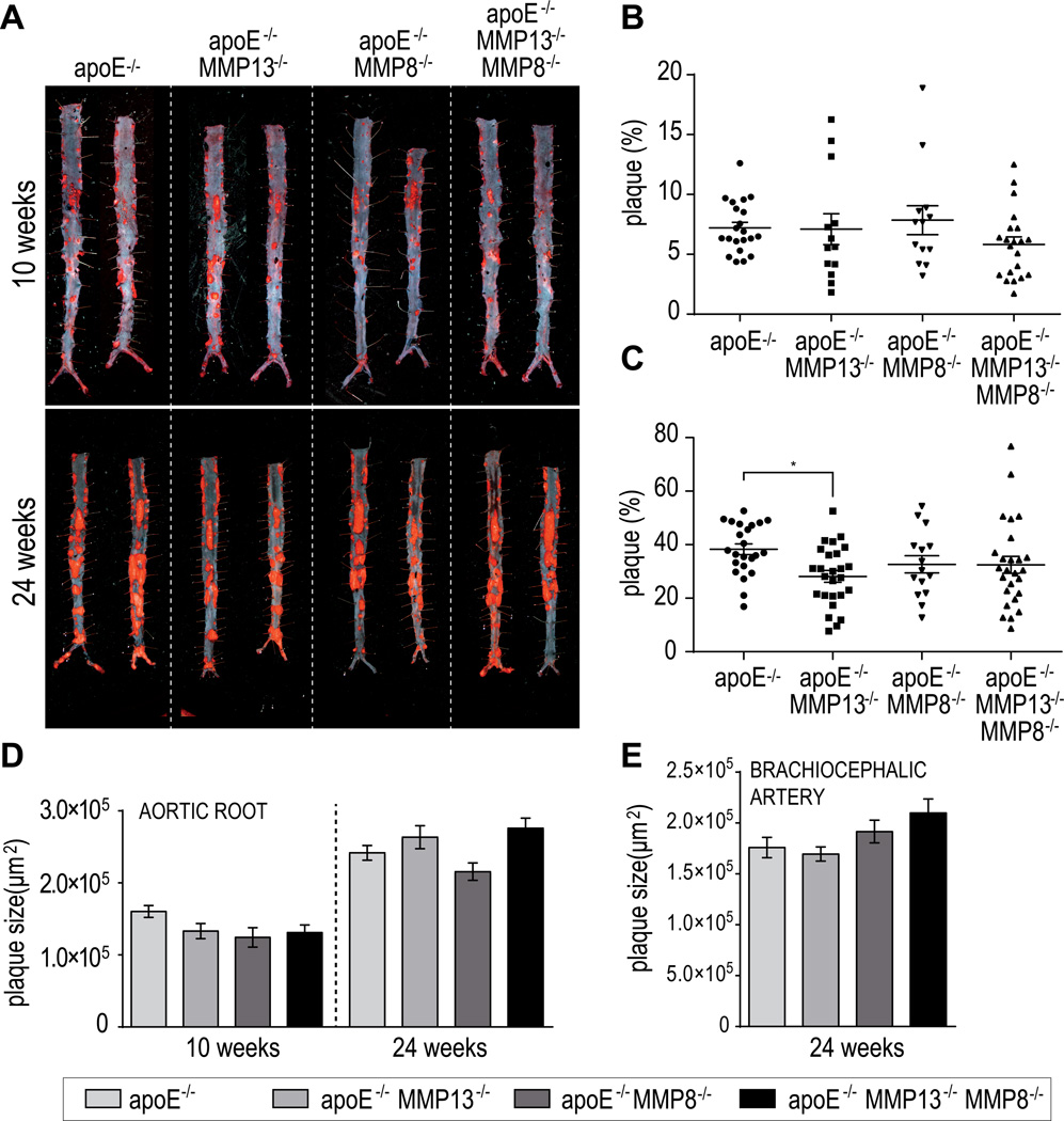
Figure 1.
Oil-red-O staining of lesions in abdominal aortas from mice after 10 and 24 weeks on an atherogenic diet (A). Quantification of positive areas after 10 weeks (B) and 24 weeks (C) on western diet. Histological analysis of plaque size on aortic root (D) and brachiocephalic artery sections. Bars represent mean ± SEM. (*p<0.05).

