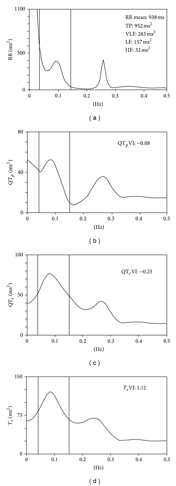
Figure 3.

Power spectral analysis of RR (a), QTe (b), QTp (c), and T e (d) calculated on a short-term (5-minute) ECG recording from the same patient shown in Figure 2. All the power spectra contain a high-frequency power component synchronous with breathing (between 0.20 and 0.30 Hz) and low-frequency power around 0.10 Hz.
