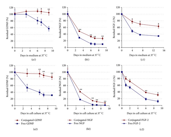
Figure 1.

Stability of free versus conjugated neurotrophic factors at 37°C in the absence ((a1), (b1), and (c1)) and in the presence ((a2), (b2), and (c2)) of cells. In the upper row free or conjugated neurotrophic factors (GDNF, βNGF, and FGF-2) were added to culture medium, each type separately (10 ng/mL, final concentration), and placed at 37°C (in the absence of cells). Aliquots were collected after different time points, and the concentration of the residual factor in the samples was measured using an appropriate ELISA kit. In the lower row, the same concentrations of free or conjugated neurotrophic factors were added once to dissociate dorsal root ganglia (DRG) cell cultures at the beginning of the experiment. The culture medium was not changed during the experiment and aliquots from it were collected at different days after cultivation. The concentration of the residual factors in the aliquots was measured as described above. The data are presented as mean ± SD in triplicate (∗P < 0.01, ∗∗P < 0.001).
