
Figure 4.
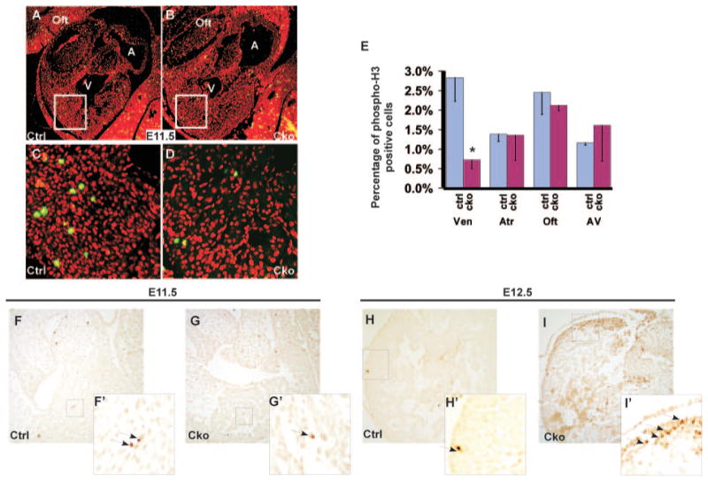
Myocardial depletion of Smad4 causes a reduction in cell proliferation and an increase in cell apoptosis. A through E, Saggital sections of control (A and C) and mutant (B and D) embryos at E11.5 were immunostained with a primary antibody against phospho-H3 (green), and total nuclei were visualized with propidium iodide staining (red). C and D correspond to the boxed regions of A and B, respectively. Quantitative analysis (E) indicates that the proliferation rate in mutant ventricles is significantly reduced compared with the control sample. At least 400 nuclei of each segment of each embryonic heart were counted. Data were averaged from 3 independent embryos with error bars indicating SD. *P<0.01 (Student t test). F through I′, TUNEL analysis was performed on sections of control (F and F′, H and H′), and mutant (G and G′, I and I′) embryonic hearts at E11.5 and at E12.5. Arrows indicate examples of TUNEL-positive cells. F′, G′, H′, and I′ correspond to the boxed regions of F, G, H, and I, respectively. A indicates atrium; Cko, cTnt-Cre;Smad4loxp/loxp; Ctrl, Smad4loxp/loxp; oft, outflow tract; V, ventricle.