Figure 1. Histological Findings in PCNSL.
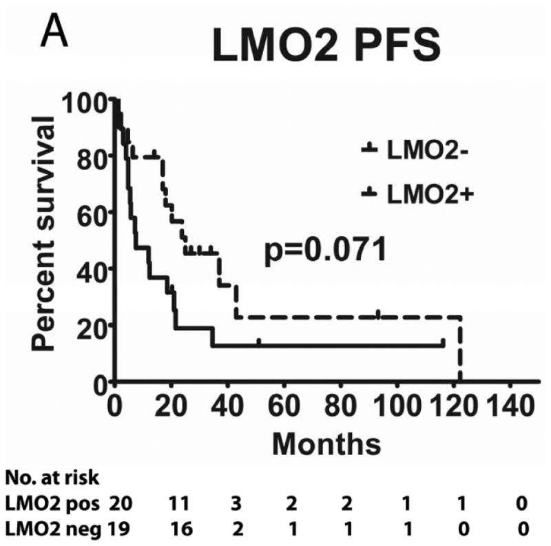
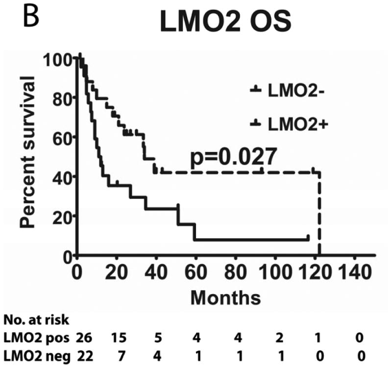
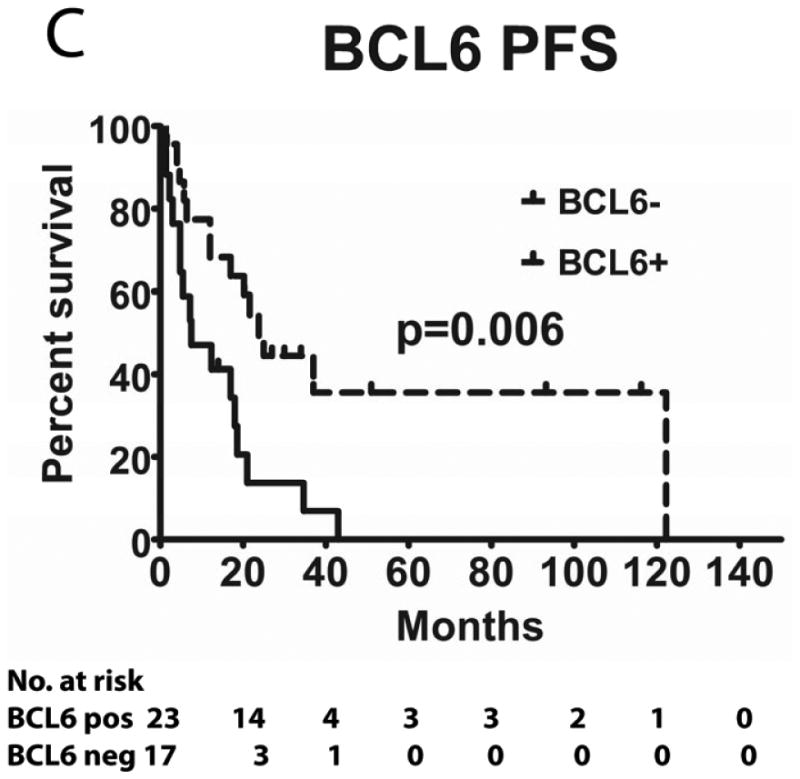
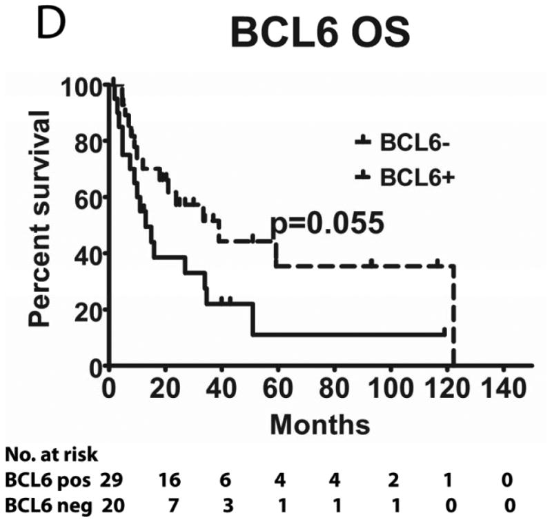
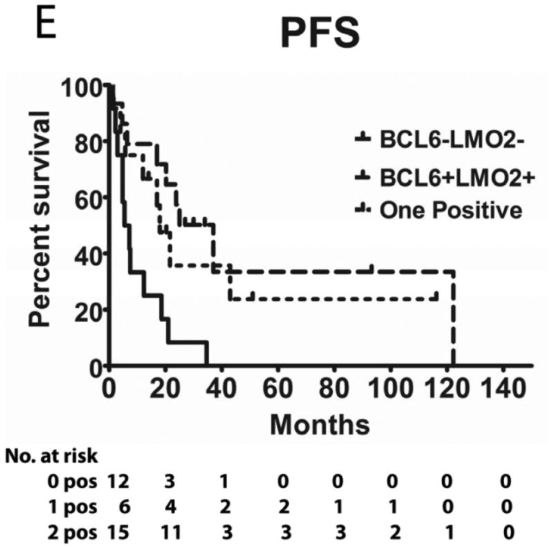
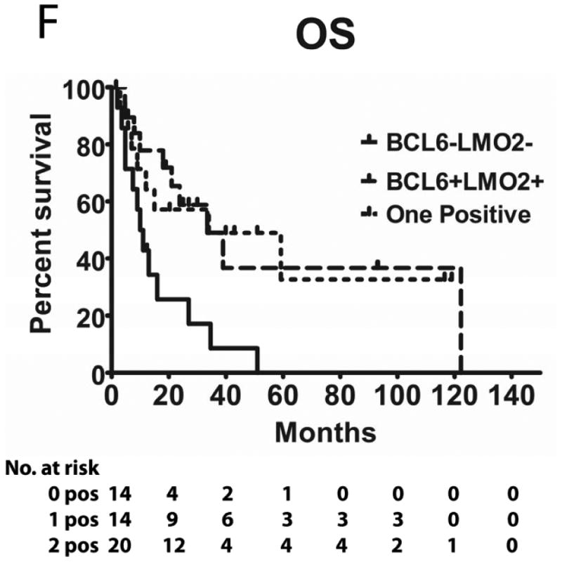
Histological sections of a representative central nervous system biopsy shows an atypical lymphoid proliferation infiltrating brain parenchyma in a diffuse pattern (A); the lymphoma cells typically surround vessels and cause angiocentric and angiodestructive lesions (B); higher magnification shows marked cytological atypia of the lymphoma cells with pleomorphic nuclear outlines, prominent nucleoli and associated mitotic figures (C); immunohistochemistry shows that the lymphoma cells are positive for HGAL (D), LMO2 (E) and BCL6 (F). [Original magnification, panel A x200, panel B x400, panels C-F x600].
