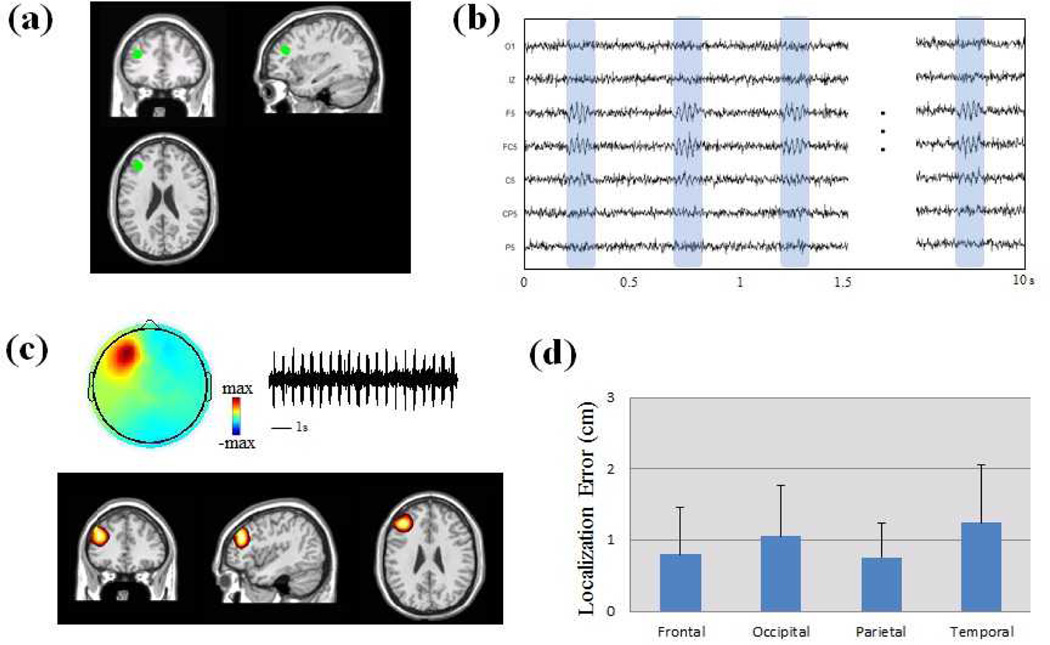
Fig. 2.
Computer simulation of HF activity in a standard head volume conduction model. (a) Location of one simulated dipole source without extent in frontal lobe. (b) Scalp EEG traces generated from the simulated source. (c) Estimated sources and independent component of the HF activity. (d) Localization error (LE) of computer simulation in 1000 trials. The simulated dipoles are categorized in four groups according to the dipole locations (Frontal, Occipital, Parietal and Temporal groups).

