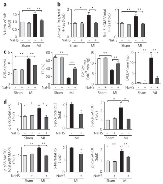
Figure 5. Electrophilic H-Ras activation regulated by HS− in cells and in vivo in cardiac tissues.
(a) Immunohistochemical determination of 8-nitro-cGMP formation in mouse hearts after myocardial infarction (MI) after NaHS treatment or in untreated (vehicle) hearts (original images are in Supplementary Fig. 8d). Data represent mean ± s.e.m. **P < 0.01. (b) Quantitative data for western blots of H-Ras activation and S-guanylation in mouse hearts (original blots are in Supplementary Fig. 8f). Data represent mean ± s.e.m. *P < 0.05, **P < 0.01. (c) Protective effects of HS− on chronic heart failure caused by myocardial infarction. Cardiac functions of mice after myocardial infarction or sham operation and treated with NaHS or untreated (treated with vehicle). LV IDd, left ventricular end-diastolic internal diameter; FS, fractional shortening; dP/dtmax, maximal rate of pressure development; EDP, end-diastolic pressure. Data represent mean ± s.e.m. **P < 0.01. (d) Quantitative data for western blots measuring amount of phosphorylation (p-) of ERK, p38 MAPK, p53 and Rb and expression of ERK, p38 MAPK, p53 and Rb in mouse hearts after myocardial infarction (original blots are in Supplementary Fig. 8f). Because western blotting detected no expression of Rb and p53 in sham-operated mouse hearts, the amounts of phosphorylation of p53 and Rb were compared after normalization with GAPDH as an internal control. Data represent mean ± s.e.m. *P < 0.05, **P < 0.01.

