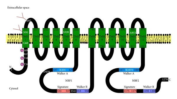
Figure 1.

Schematic model of ABCB4. ABCB4 consists of twelve TMHs spanning the plasma membrane and two cytosolic NBFs containing the Walker A, Walker B, and signature motifs. N and C indicate the N- and C-termini of ABCB4, respectively. TMHs are predicted from the crystal structure of mouse Abcb1a (PDB code 3G61) reported by Aller et al. [60]. The residues N91 and N97 are N-glycosylated with complex oligosaccharides. The residues T34, T44, and S49 are phosphorylation sites. The T34M, R47G, K435M, and K1075M mutations impair the phospholipid efflux activity of ABCB4. The G68H, S320F, D459H, I541F, and A953D mutations result in the intracellular retention of ABCB4.
