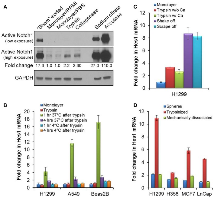
Figure 1.
Activation of Notch1 after detachment in cell culture. (A) The cleaved, activated form of Notch1 was blotted (Cell signaling #4147 anti-Notch1 Val1744 antibody) on lysates from H1299 cells immediately after sorting (“sham”-sorted), from monolayer cells after a brief rinse in RPMI media or PBS, or from cells detached with trypsin, collagenase, sodium citrate, or accutase. (B) Quantitative RT-PCR of Hes1 from mRNA samples collected either from attached monolayer cells immediately after trypsinization, or after an incubation in complete media at 37°C, 5% CO2, or on ice for 1 or 4 h after trypsinization. (C) Quantitative RT-PCR of Hes1 from mRNA collected from attached H1299 monolayer cells, or after incubation of H1299 cells at 37°C, 5% CO2 for 1 h after cell detachment by trypsin without (w/o) or with (w/) 0.5 mM CaCl2, by gentle agitation (shake off) or by cell-scraping (scrape off). (D) Quantitative RT-PCR of Hes1 from mRNA samples collected from tumorspheres or from dissociated tumorsphere cells that were incubated at 37°C, 5% CO2 for 1 h after dissociation either enzymatically by trypsinization or mechanically by pipetting. Error bars represent standard deviation.

