
Figure 1.
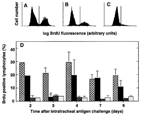
Flow cytometric analysis of in vivo lymphocyte proliferation. SRBC-primed C57BL/6 mice were challenged intratracheally with SRBC to induce a pulmonary immune response. On various days after challenge, BrdU (4 mgs/mouse IP) was administered in 1-3 doses at 20 minute intervals before the mouse was killed and cells harvested by BAL and dissection. Cells were stained for incorporated BrdU as described in Methods and were analyzed by flow cytometry. A-C. Representative histograms of cells within light scatter-defined gates from (A) bone marrow, (B) PTN, and (C) BAL. Data are from a single mouse four days after intratracheal antigen challenge. D. Kinetics of BrdU incorporation. Bars represent BrdU+ cells (defined by light scatter gating) in bone marrow (light cross hatching); paratracheal nodes (black); BAL (dark cross-hatching); and lung mince (light stippling). Data are mean ± SEM of 3 experiments, each consisting of pooled samples from 2-5 mice (except for bone marrows, which were assayed individually). *, significantly different from same tissue at other time-points, p <0.05, ANOVA with Fisher's PLSD post hoc testing.