
Figure 8.
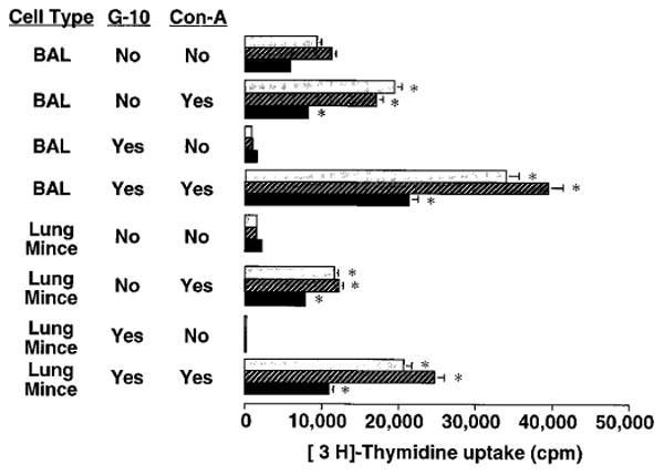
Proliferative response of lung lymphocytes to Con-A stimulation in vitro. BAL and lung mince mononuclear cells were harvested from SRBC-primed C57BL/6 mice four days after induction of a pulmonary immune response by IT challenge. Aliquots were depleted of phagocytic cells including macrophages by Sephadex G10 column. Cells were cultured for 2 days (light stippled bars), 3 days (cross-hatched bars), or 4 days (black bars) at 2 × 10⁵ cells per well in flat-bottomed 96-well plates in complete medium in the absence or presence of Con-A 10 μg/mL (an optimal dose). Lymphocyte proliferation was assessed by uptake of 3H-Thd during the final 16 hours of culture. Splenocytes in the same experiment gave 18,000-98,000 cpm. *, significantly different compared to corresponding condition without Con-A stimulation, p < 0.05, unpaired t test. Similar results were obtained in two separate experiments.