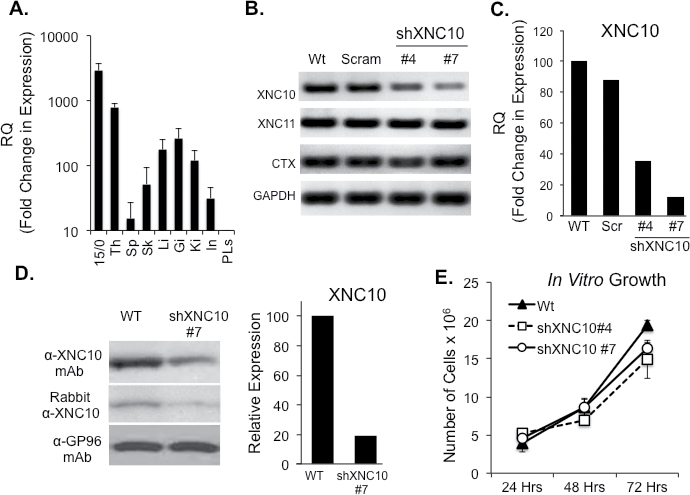
Fig. 1.
XNC10 expression pattern and generation of stable XNC10-deficient 15/0 tumor cell lines. (A) XNC10 gene expression in normal tadpoles tissues and 15/0 tumor cells by qPCR. Th, thymus; Sp, spleen; Sk, skin; Li, liver; Gi, gills; Ki, kidney; In, intestine; PLs, peritoneal leukocytes. (B) 15/0 tumor cells were transfected with either a scrambled shRNA or an anti-XNC10 shRNA construct and stable clones were generated. RT–PCR gene expression analysis of XNC10, XNC11, CTX and GAPDH in 15/0 WT, shScramble, shXNC10 clone #4 and #7 tumor cells. (C) Quantitative RT–PCR gene expression analysis of XNC10 on 15/0 WT, shScramble, shXNC10 clone #4 and #7 tumor cells. (D) Western blot analysis of anti-XNC10 antibodies. Left: 15/0 WT and shXNC10#7 cell lysates incubated with the 291 monoclonal antibody (top) or rabbit polyclonal (bottom) anti-XNC10 antibodies. Left: Densitometry analysis of the XNC10 protein expression assessed using the 291 monoclonal antibody. (E) Assessment of tumor transfectants’ proliferation; 1×10⁵ cells of 15/0 WT, shXNC10 #4 or shXNC10 # were plated in triplicate and grown in vitro then cells were collected and counted 24, 48 and 72h later. n = 3 independent experiments.

