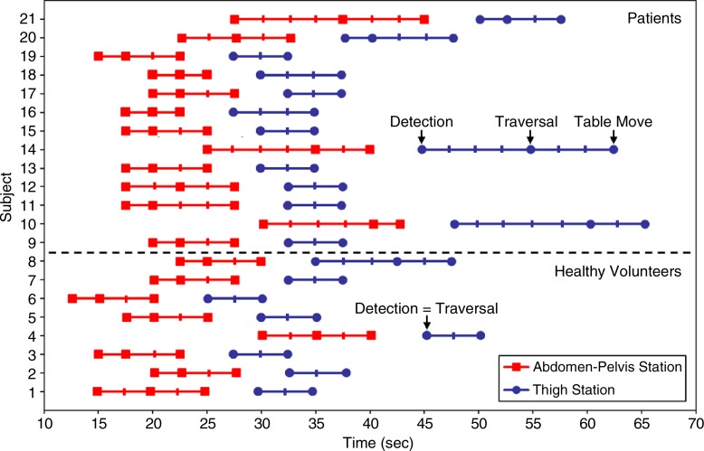
Figure 2:
Graph shows contrast material bolus detection for all 21 subjects. In many cases, bolus had already traversed thigh station by the time acquisition began there; thus, bolus detection and traversal times were the same. Subjects 1–8 were healthy volunteers and subjects 9–21 were patients. Hash marks on the subject lines indicate fluoroscopic tracking time frame intervals (2.5 seconds), and times are relative to start of contrast material administration.

