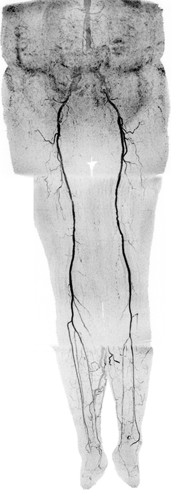
Figure 6:

Coronal MIP in a 73-year-old female patient (subject 12) with extensive motion-related artifacts at abdomen-pelvis station and rapid arterial-to-venous transit at calf-foot station. Because of these effects, CT angiography was scored marginally better. Angiogram consists of final abdomen-pelvis and thigh station time frames and third calf-foot time frame. Also see Movie 4 (online).
