
Figure 2.
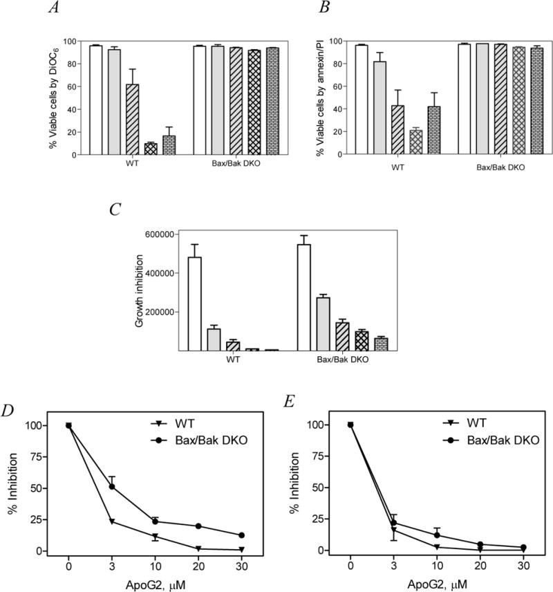
Bax and Bak expressing (WT) and double knockout (DKO) mouse embryo fibroblasts were incubated with 0 – 30 μM of apogossypolone and the viability of cells was tested by two standard apoptosis assays. A. DiOC6 method. B. Annexin/PI binding assay. Growth inhibition was measured by cell count method. C. Bar graph representing decline in total number of cells at the end of 24 hrs incubation with apogossypolone. D and E. Line graph representing dose dependent percent decrease in cell number upon incubation with apogossypolone for 24 hrs (D) or 48 hrs (E). All experiments were representatives of triplicates and the data are mean ±SEM of three similar sets of experiments.