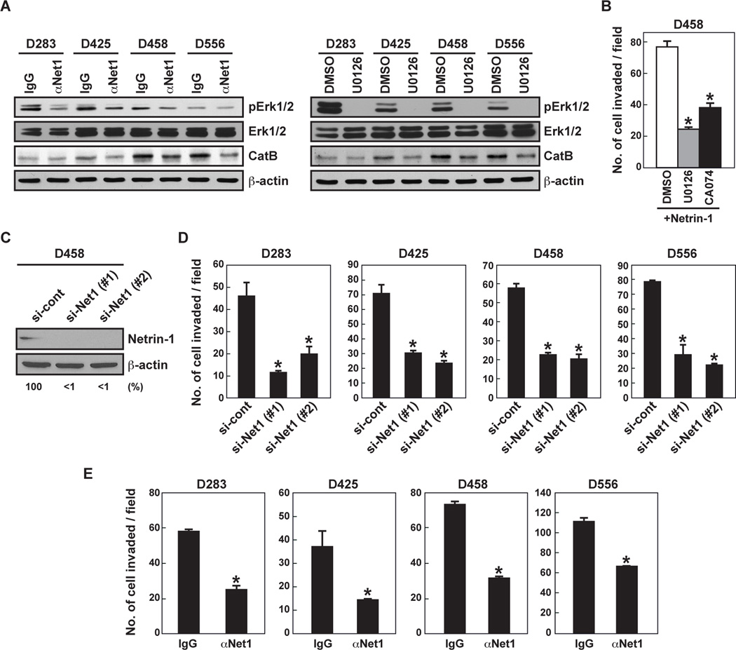
Figure 2. Inhibition of netrin-1 blocks MB cell invasion and Erk phosphorylation.
A, MB cells were treated with netrin-1 neutralizing antibody (20 µg/mL, for 120 minutes) or U0126 (10 µM, for 30 minutes) prior to cell lysate collection. Cell lysates were analyzed by western blot. B, D458 cells were incubated with MEK inhibitor (U0126, 10 µM) or CatB inhibitor (CA074, 10 µM) and cell invasiveness was assessed with Matrigel-coated Transwells. C, D458 cells were transfected with control or netrin-1-specific siRNA (#1 and #2) (20 nM). After 24 hours, the silencing effect of netrin-1 siRNA on netrin-1 protein levels was analyzed by western blot. The intensity of netrin-1 bands was normalized to their respective β-actin controls. Numbers below gel lanes represent the fold-change in intensity relative to controls. D, MB cells were transfected by netrin-1 siRNA and invasion assay was performed. Data represent the mean ± SD (n=3), * p < 0.05. E, MB cells were incubated with control IgG or netrin-1 neutralizing antibody (20 µg/mL). Cell invasiveness was assessed via Matrigel-coated Transwells. Data represent the mean ± SD (n=3), * p < 0.05.

