
Figure 3. Extended appearance of microglia/macriphage in the subretinal space in Ccl3-/-Abca4-/-Rdh8-/- mice after light exposure.
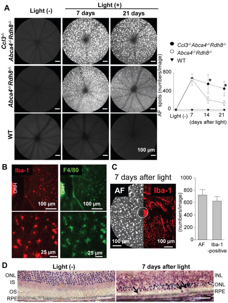
Ccl3-/-Abca4-/-Rdh8-/-, Abca4-/-Rdh8-/- and WT mice at 4-6-week-old age were exposed to 10,000 lux light for 30 min. A. Retinal images were captured by in vivo SLO. Images were taken at 7, 14, and 21 days after light exposure (left). Bars indicate 100 μm. Numbers of autofluorescent (AF) spots of each image were counted (right). Error bars indicate S.D. of the means (n > 6).* indicates P < 0.05 vs light exposed Abca4-/-Rdh8-/- mice. B. RPE flat-mounts of Abca4-/-Rdh8-/- mice were prepared 7 days after light exposure and stained with anti-Iba-1 (left) and anti-F4/80 (right) Abs. Lower panels show magnified images. ONH, optic nerve head. C. SLO image (left) and flat-mount IHC with anti-Iba-1 Ab (right) in the same magnification is presented. Abca4-/-Rdh8-/- mice were exposed to 10,000 lux for 30 min and kept in the dark for 7 days. Numbers of AF spots in SLO images (per image) and Iba-1-positive cells in IHC in the same size area were counted (right graph). ONH was circled by broken line. D. Retinal cross section of Abca4-/-Rdh8-/- mice without light exposure and 7 days after light exposure at 10,000 lux for 30 min was prepared by Epon-embedment. Arrows indicate cells in the subretinal space. Bars indicate 50 μm. ONL, outer nuclear layer; IS, inner segment; OS, outer segment; INL, inner nuclear layer.