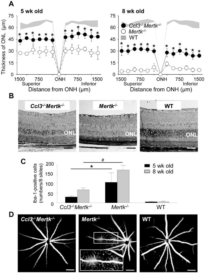
Figure 8. Ccl3 deficiency protects the retina from degeneration in the mouse model for retinitis pigmentosa.

Ccl3-/-Mertk-/- mice were established by crossing between Mertk-/- and Ccl3-/- mice. Littermate Mertk-/- mice were used as control. A. Thickness of ONL from Ccl3-/-Mertk-/-, Mertk-/- and WT mice at 5-week and 8-week of age were measured by SD-OCT. Error bars indicate S.D. of the means (n > 6). * indicates P < 0.05 vs Mertk-/- mice. B. Images of retinal histology from 5-week-old Ccl3-/-Mertk-/-, Mertk-/- and WT mice are shown. Bars indicate 50 μm. ONL, outer nuclear layer. C. Iba-1-positive cells in the subretinal space were counted using cryosections from Ccl3-/-Mertk-/-, Mertk-/- and WT mice at 5-week and 8-week of age. These cryosections were prepared every 200 μm distance from the edge to edge (8 slides/eye), and IHC was performed with anti-Iba-1 Ab. Error bars indicate S.D. of the means (n > 6) *, # indicates P < 0.05. D. Fluorescent angiography from 8-week-old Ccl3-/-Mertk-/-, Mertk-/- and WT mice are presented. Magnified images of solid line inset shown in the broken rectangle.
