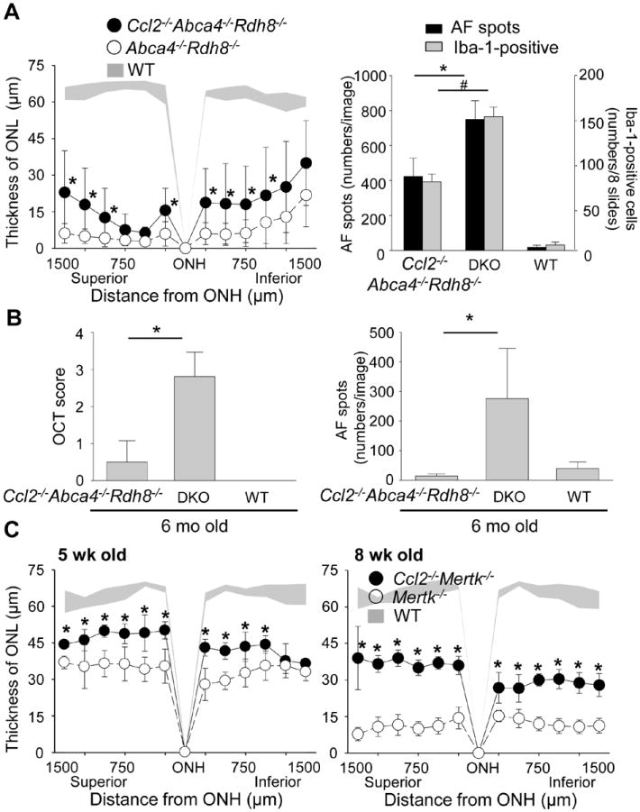
Figure 9. Ccl2 deficiency protects the retina from degeneration in Abca4-/-Rdh8-/- mice and Mertk-/- mice.

A. Ccl2-/-Abca4-/-Rdh8-/-, Abca4-/-Rdh8-/- (DKO) and WT mice at 4-6-week-old age were exposed to 10,000 lux light for 30 min. Thickness of ONL was measured by SD-OCT at 7 days after light exposure (left). Error bars indicate S.D. of the means (n > 6).* indicates P < 0.05 vs light exposed Abca4-/-Rdh8-/- mice. Numbers of AF spots of each image of SLO and Iba-1-postitive cells in the subretinal space were counted 7 days after light exposure (right). Cryosections were prepared every 200 μm distance from the edge to edge (8 slides/eye), and IHC was performed with anti-Iba-1 Ab. Error bars indicate S.D. of the means (n > 6).*, # indicates P < 0.05. B. Ccl2-/-Abca4-/-Rdh8-/-, Abca4-/-Rdh8-/- (DKO) and WT were kept under regular light conditions (12h ~10 lux/12 h dark), and retinal phenotype of these mice were characterized at the age of 6 month. Severity of retinal degeneration was evaluated by established scoring system (20) with in vivo OCT imaging (left). * indicates P < 0.05. Numbers of AF spots was counted by in vivo SLO imaging (right). * indicates P < 0.05. C. Thickness of ONL from Ccl2-/-Mertk-/-, Mertk-/- and WT mice at 5-week and 8-week old age were measured by SD-OCT. Error bars indicate S.D. of the means (n > 6). * indicates P < 0.05 vs Mertk-/- mice.
