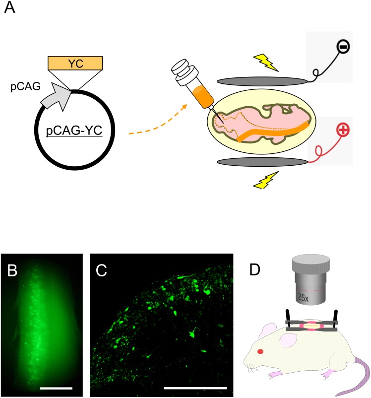
Figure 1. Methodological overview.
(A) In utero electroporation of Yellow Cameleon expression vector. pCAG-YCnano50 was injected into the spinal cord central canal at E12.5 and electric pulses were applied with an electroporator. (B) YC expression in the spinal dorsal horn of an 8-week-old mouse. YC was expressed in the left side of the spinal cord around the L1 level. Scale bar, 1 mm. (C) Transverse section of YC-expressing SDH at L1. SDH on the left side is shown. Scale bar, 200 µm. (D) In vivo calcium imaging of SDH neurons was performed in YC-expressing mice (8–10 weeks old). The spinal cord at the level of L1 was exposed by laminectomy. The mouse was fixed in a stereotactic frame by attaching custom-made clamps to the vertebral column, and calcium imaging was performed by use of a two-photon microscope.

