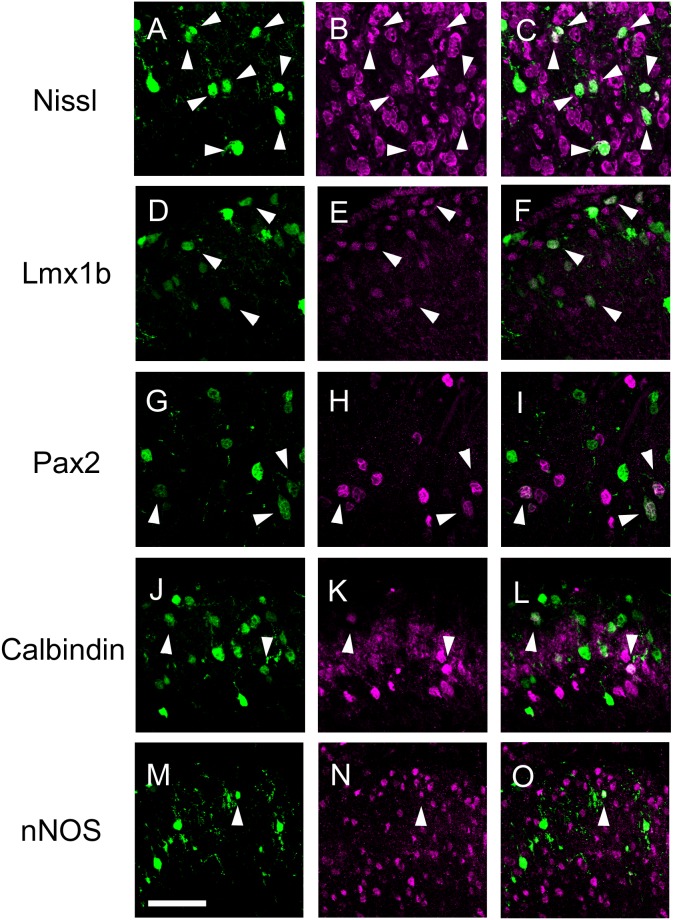
Figure 2. Identity of EGFP-expressing cells in the SDH.
pCAG-EGFP was electroporated into the spinal cord at E12.5, and transverse sections were prepared from the thoracic spinal cord of the electroporated mice at P21. The sections were Nissl-stained (B, C) or immunostained with anti-Lmx1b (E, F), anti-Pax2 antibody (H, I), anti-Calbindin antibody (K, L), or anti-nNOS antibody (N, O). EGFP fluorescence (A, C, D, F, G, I, J, L, M, O), and immunofluorescence of anti-Lmx1b (E, F), anti-Pax2 (H, I), anti-Calbindin (K, L), and anti-nNOS (N, O) are shown. Arrowheads indicate double-positive cells. Scale bar, 50 µm.

