
Figure 4.
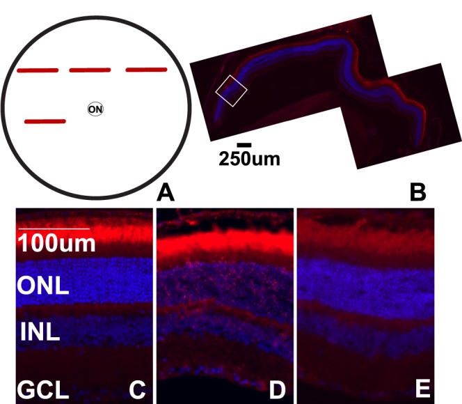
Cell death post blast is focal. (A) Schematic of a flat-mount view of the retina, red bars indicate regions within retinal cross-sections that are TUNEL-positive. (B) Epifluorescence micrograph of a TUNEL-positive midperipheral retinal cross-section from a 28-day postblast eye. The white box indicates an area of TUNEL-positive cells. (C–E) Representative epifluorescence micrographs of retinas from control (C), or 28-day postblast retinas labeled with TUNEL (red) and DAPI (blue). Affected (D) and unaffected (E) retinal regions from the same blast eye are shown. ON, optic nerve.