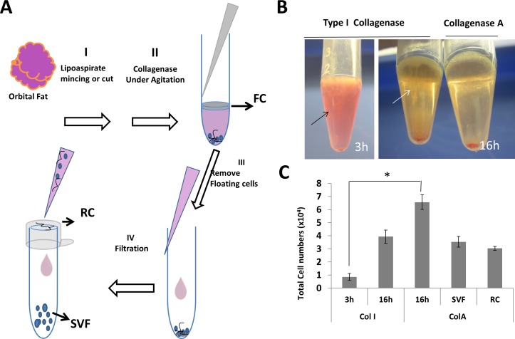
Figure 1.
Different cell fractions from human orbital adipose tissue. After surgical removal, human orbital adipose tissues were cut in smaller pieces (Step I), subjected to collagenase digestion (Step II), centrifuged to remove FCs (Step III), and filtered to obtain SVF in the flow through and RC from cells caught on the filter. (Step IV) (A). Different appearances were noted after centrifugation following digestion with Col I for 3 and 16 hours, and with Col A for 16 hours (B). Such a difference was reflected by the total cell number of cells in the pellet after digestion in Col I or ColA for 3 or 16 hours, as well as SVF and RC following Step IV filtration (C).

