
Figure 7. Dye release normalized to the change in tryptophan fluorescence quenching measured for β2m with BMP-containing LUVs at pH 4.5.
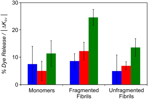
The ratio of % dye release per membrane interaction detected via a change in tryptophan quenching observed in the absence or presence of lipid. β2m monomers, fragmented and unfragmented fibrils (6 µM monomer equivalent concentration) were incubated in Assay Buffer pH 4.5 with LUVs (5 µM lipid) comprising 36 POPC: 20 POPE: 7 SM: 25 cholesterol (mol/mol) plus 0 mol % (blue), 12 mol % (red) or 50 mol % (green) BMP for 10 min at 37°C. Error bar represent 1 S.E.