Fig. 7.

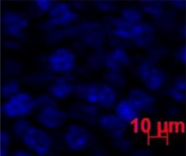
Representative fluorescence image of the cornea at 6 h after subconjunctival injection of Alexa647. Blue color represents the cell nucleus stained by DAPI. No red color that represents the Alexa647 marker could be found in the image.

Representative fluorescence image of the cornea at 6 h after subconjunctival injection of Alexa647. Blue color represents the cell nucleus stained by DAPI. No red color that represents the Alexa647 marker could be found in the image.