
FIGURE 1.
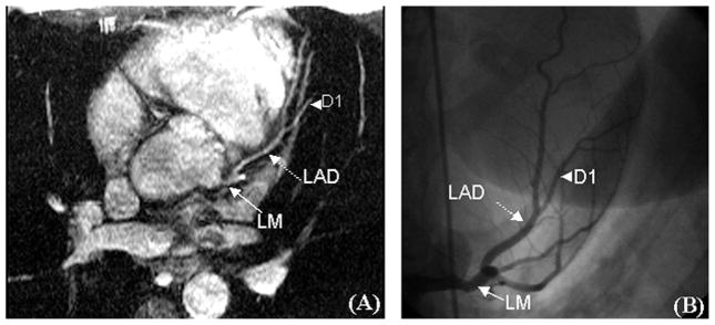
Coronary angiograms of the LM and LAD in a 45-year-old with palpitations and a positive stress echocardiogram. A, The MRA image of the normal LM and LAD. B, The corresponding normal x-ray angiogram. D1 is the first diagonal branch of the LAD (arrowhead).