
FIGURE 3.
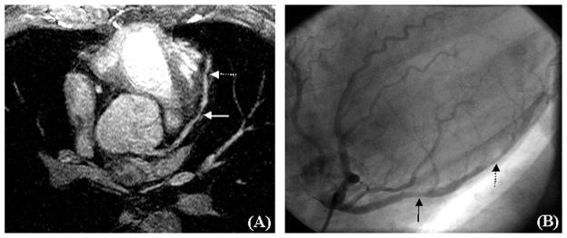
Coronary angiograms of the LM and LAD in a 54-year-old asymptomatic male marathon runner who was referred for further evaluation of a high calcium score detected on screening computed tomography. A, The MRA image, and B, The x-ray angiogram. There is a 30% stenosis in the proximal LAD (solid arrows) and a 60% stenosis in the mid LAD (broken arrows).