
Fig. 7.
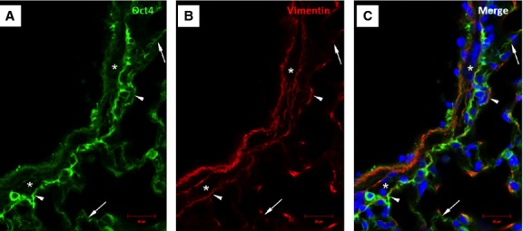
Immunofluorescence of Oct4 and vimentin in the vascular wall and perivascular regions. (A) Oct4-positive cells circumscribe the smooth muscle cell layer (asterisk). (B) Immunolabeling for vimentin. (C) Arrowheads in the merged picture indicate cells which are positive for both, Oct4 and vimentin. Note that Oct4-positive cells extend to the alveolar septum (arrows); bar = 10 μm.