
Fig. 8.
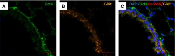
Immunofluorescent confocal images of Oct4 and C-kit labelling pattern in bronchus. (A) Oct4-positive cells are present in the peribronchial area. (B) Some epithelial cells are positive for c-kit. (C) The merged picture demonstrates little co-localization of Oct4 with C-kit. α-SMA labelling (red) of smooth muscles lining the bronchiolar epithelium; bar = 10 μm.