
Fig. 14.
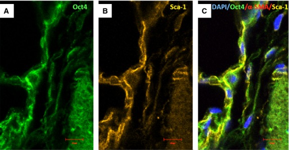
Immunofluorescence of Oct-4 and Sca-1 in blood vessels. (A) Oct4-positive cells are present in the perivascular areas and in the vascular wall. (B) Sca-1 is also expressed in such areas. (C) The merged picture demonstrates co-localization of Oct4 and Sca-1. α-SMA (red) labels vascular smooth muscle cells (C); bar = 10 μm.