
Fig. 15.
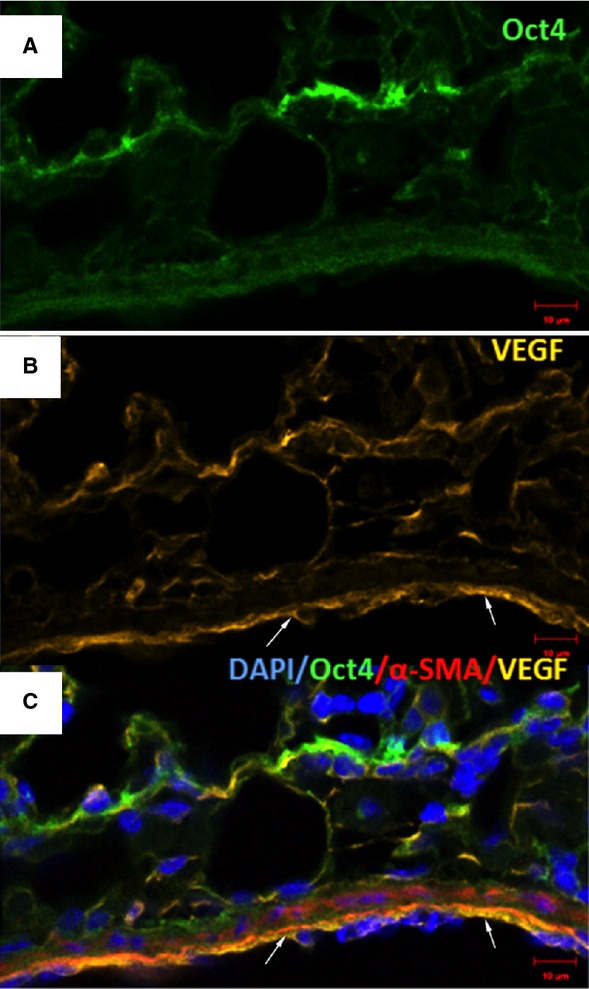
Immunofluorescence of Oct4 and VEGF in blood vessels. (A) Oct4-positive cells are located in the perivascular areas and co-localize with VEGF (B). Notice abundant VEGF expression in endothelial cells (arrows). (C) The merged picture demonstrates co-localization of Oct4 and VEGF. α-SMA (red) labels smooth muscle cells in the media; bar = 10 μm.