
Fig. 1.
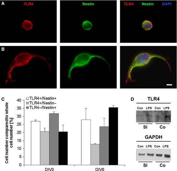
Toll-like receptor 4 (TLR4) receptors in the small intestine. Immunofluorescent staining TLR4 (red), nestin (green) and DAPI (blue) of fresh isolated (A) and cultivated (B) Neural stem/progenitor cells; bar: 5 μm. Quantification of the amount of TLR4-positive and nestin-positive cells (C). The cells, which are neither TLR- nor Nestin-positive, are represented in the black column. Western blot analysis indicated the presence of TLR4 protein in small intestine and colon with and without lipopolysaccharides Stimulation (loading control: GAPDH; D).