
Fig. 1.
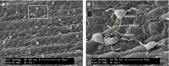
Distribution of vascular telocytes (TC) in pig, scanning electron microscope. (A) Under lower power, several TC are observed on the endothelial surface. (B) Local enlargement of white rectangle of A indicates a typical TC with triangular cell body and one long Telopode (Tps) (Tp, 11.9 μm in length) with alternation of podomers and podoms.