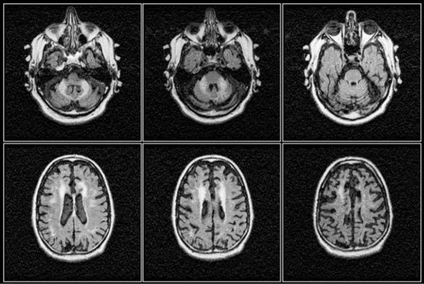
Figure 2.
Axial FLAIR images of the brain of the 64 year old male carrier of 80 CGG repeats with typical manifestations of FXTAS comprising marked cerebellar ataxia, intention tremor, autonomic failure (originally diagnosed with multiple system atrophy, MSA). Marked bilateral T2 hyperintense signals within the middle cerebellar peduncles (MCP sign), combined with symmetrical T2 hyperintense signals within the corona radiate and considerable cerebral and cerebellar atrophy are evident from the images.166 Reproduced from Loesch DZ et al. Clinical Genetics 2005; 67(5):412-417.

