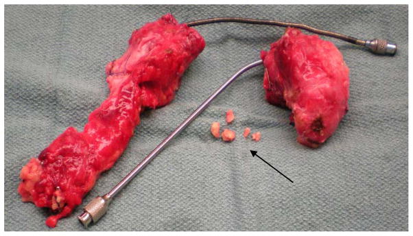
Figure 2.

Chronic pancreatitis pancreas resected from male pediatric donor at age 12. Noted were severe fibrosis (white tissue color), rock-like texture, heavy blood infiltration, and medium level of parenchymal fat. Depicted is the pancreas split into head and body/tail sections with large metal catheters used during surgical cannulation to accommodate the dilated main pancreatic duct. Also, several calcification deposits (arrow) up to 3mm in diameter were found intraductally in the head region.
